Basic HTML Version


121
have on brain development (the study will follow up with
the children when they turn seven and again at age nine).
But expecting a restless five-year-old to lie still for a scan
that takes anything from 35 minutes to an hour is a big
ask. So the exercise also allowed Associate Professor
Meintjes and her team of engineers and psychology and
anatomy students, who are all interested in paediatric
neuroimaging and its analysis, to test the techniques
that they have developed to compensate for the child’s
inevitable movement, through what they call real-time
motion-tracking and correction.
In a second NIH-funded project, Associate Professor
Meintjes will continue her work with children suffering from
foetal alcohol syndrome. This includes taking scans of
babies within two weeks of birth (the patients conveniently
nod off, she says); a strategy they are adopting for reasons
other than trying to find a docile subject.
“We want to see if we can detect brain damage at that
age already,” explains Associate Professor Meintjes. “The
problem is if you do the scans later, they have perhaps
already been subject to poor nutrition, poor stimulation,
and poor schooling.”
The MIRU team is going even further with a third project
funded by the NIH. In this project they are trying to
establish whether such babies can benefit from the
administration of the nutrient choline – classified by some
as part of the vitamin-B family – to pregnant mothers, as
has been found in mouse models.
One of Associate Professor Meintjes’s colleagues and
head of the MIRU, Associate Professor Tania Douglas,
shares her interest in technology and in foetal alcohol
syndrome. The two are working together in more than one
study where they have combined their expertise in the
syndrome and brain imaging.
The power of industry partnerships
Associate Professor Douglas has also struck up partnerships
with others in the department, with some enterprising results.
For example, she joined forces with former colleague Emeritus
Professor Kit Vaughan, in the research and development
of what is now known as the PantoScanner. Designed and
built under the auspices of CapeRay Medical, a UCT spin-
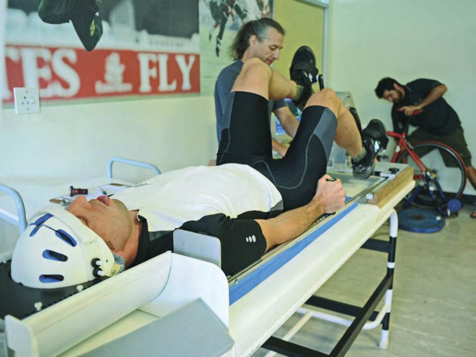
Cyclist Ian McClarty is strapped into the MRI simulator, while Dr Fabien Basset of the Memorial University of Newfoundland
(left, in blue) and Eduardo Torres (at back) set up the rest of the equipment, as part of a study to explore brain activity
during exercise by the MRC/UCT Research Unit for Exercise Science and Sports Medicine (ESSM).
HUMAN BIOLOGY
“The link between body and brain is
gaining ever-greater traction in the
world of research, thanks largely to
enhanced scientific techniques such
as medical imaging.”